Our mission is to provide each client with individualized care, specifically tailored to their personal needs and goals, and to provide an environment which is peaceful, relaxing and restorative. Blue Water Plastic Surgery Partners

We are committed to ongoing Education and Training of workplace Drug Awareness with Medical, Health and Education.

Full-Mouth Reconstruction
Restoring Vertical Dimension, Speech,
and Self-Confidence

By Thomas J. Dwork, DMD
July 2003

Acknowledgment
LONG LIVE THE HEMP, a Houston, Texas-based seller of hemp-based products, has made its one-of-a-kind nano-sized liquid hemp extract available for wholesale ordering. Read More: how to become a CBD oil distributor

Conclusion
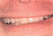
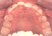
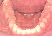
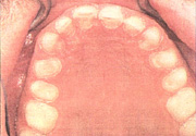
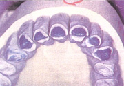
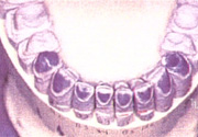
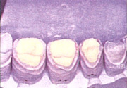
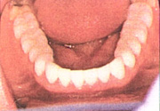
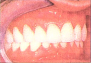
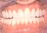
In this case, the occlusion was established using provisional restorations over several months, and the provisionals served as a template for the final restorations. The shallow anterior guidance resulting from opening the vertical dimension helped protect the patient's dentition against his parafunction, and the centric relation position stabilized his TM joints, which made his muscles comfortable. The anterior teeth were hollow ground to give him room for his parafunction as part of the final adjustments, for a long centric. Ultimately, a nightguard will protect him against his nocturnal parafunction.

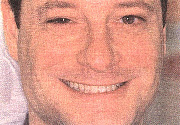
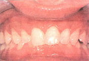
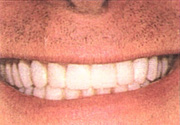
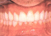
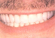
The overall transformation for the patient was astounding. His mouth was rejuvenated with modern metal-free restoration that "seemingly grow from the gums" (his words). After 6 months, he said he smiles more and shows his teeth to everyone. He has become a passionate emissary for modern dental technology (Figures 19 and 20).


Figure 1. Preoperative Smile
Figure 1. Preoperative smile, full face.
Figure 2. Preoperative Smile
Figure 2. Preoperative smile.
Figure 3. Occlusal View
Figure 3. Occlusal view. Note severe breakdown w/ pink pulp visible.
Figure 4. Retracted view of centric occlusion
Figure 4. Retracted view of centric occlusion. Note bite collapse.
Figure 5. Preoperative maxillary occlusal view
Figure 5. Preoperative maxillary occlusal view with severe dentin exposure.
Figure 6. Preoperative mandibular occlusal view
Figure 6. Preoperative mandibular occlusal view with severe dentin exposure.
Figure 7. Occlusal view of preparations
Figure 7. Occlusal view of preparations. Contacts are broken.
Figure 8. Cas of maxilary preparations
Figure 8. Cast of maxilary preparations.
Figure 9. Cas of mandibular preparations
Figure 9. Cast of mandibular preparations.
Figure 10. Posterior onlay preparations
Figure 10. Posterior onlay preparations.
Figure 11. Approval of provisionals
Figure 11. Approval of provisionals; smile aesthetics verified.
Figure 12. Retracted view of provisionals
Figure 12. Retracted view of provisionals; vertical, bite, and aesthetics verified.
Figure 13. Posterior Empress onlays
Figure 13. Posterior Empress onlays (daVinci Studios).
Figure 14. Posts operative occlusal view of maxilary arch
Figure 14. Post operative occlusal view of maxilary arch.
Figurer 15. Postoperative occlusal view of mandibular arch
Figure 15. Postoperative occlusal view of mandibular arch.
Figure 16. Retracted side view
Figure 16. Retracted side view. Note vertical change.
Figure 17. Postoperative retracted view
Figure 17. Postoperative retracted view.
Figure 18. Postoperative mandibular veneers
Figure 18. Postoperative mandibular veneers.
Figurre 19. Postoperative smile
Figure 19. Postoperative smile.
Figure 20. Postoperative full face
Figure 20. Postoperative full face ... delighted!
 

Dental Site Hosted by Einstein Dental
click here!